Retinoschisis
Acquired Retinoschisis
The retina consists of many neural layers
and as we age
sometimes these layers can split into two.
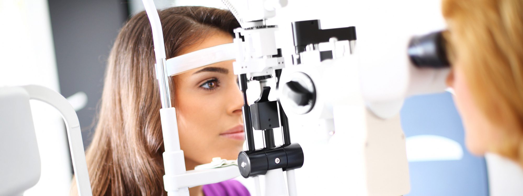
The retina is the light-sensitive tissue that lines the back of the eyeball
sending visual impulses through the optic nerve to the brain. It acts like film in a camera
capturing everything we see and transmitting it to the brain for interpretation. The retina consists of many neural layers
and as we age
sometimes these layers can split into two. This is known as acquired retinoschisis and the condition affects about five percent of the population. The vast majority of cases are harmless but the area of the retina affected usually has reduced sensitivity to light (visual field defect). Since the splitting typically occurs in the extreme periphery of the retina
most people are unaware of any vision loss.
Retinoschisis is not the same as a retinal detachment. A retinal detachment involves all layers of the retina separating from the back of the eye and is often more visually threatening than retinoschisis.
There are two types of retinoschisis: juvenile (congenital) and acquired. The juvenile form is a rare inherited condition that only affects males and has the potential to be more vision-threatening. Acquired retinoschisis is much more common and is usually seen in both males and females after age 20 and on
but most commonly after age 40. The majority of patients are hyperopic (farsighted)
and the condition affects both eyes in more than half the cases.
Fortunately
very few people with acquired retinoschisis have vision loss and no treatment is warranted. In certain situations
however
the separating of the layers threatens the macula
the part of the retina responsible for central vision. In this case
surgery or laser therapy may be performed to prevent further progression and vision loss. Moreover
in rare cases retinoschisis can progress into a retinal detachment. Due to these risks
routine retinal examinations are suggested so that changes in the retina may be monitored.