High Blood Pressure and How it Effects Vision
Contents |
High Blood Pressure and How it Effects Vision
High blood pressure, known medically as hypertension, can cause many different complications in the body, including changes in the circulation within the eye. Hypertension is often called “the silent killer” because there are usually no outward signs or symptoms that would alert an individual to its presence.

The pressure within the blood vessels must be enough that all bodily tissues have uncompromised circulation. However, when blood pressure becomes too high, it can damage artery walls and cause organ failure if left untreated.
A blood pressure reading looks like a fraction, with one number above another, like this: 120/85. The first number is the pressure in the arteries, as the blood is being pumped outward, called systolic, while the second is the venous pressure, as it is returned to the heart, called diastolic. Recent guidelines indicate that someone with systolic pressure above 140 and/or diastolic pressure above 90 should be treated for hypertension with lifestyle changes, diet, medication or a combination of these.
Increased pressure in the blood vessels makes the heart work much harder to force the blood, carrying oxygen and nutrients into all areas of the body. Over time, the increased pressure causes damage to the arteries by thickening their walls and making these walls harder, a condition called arteriosclerosis.
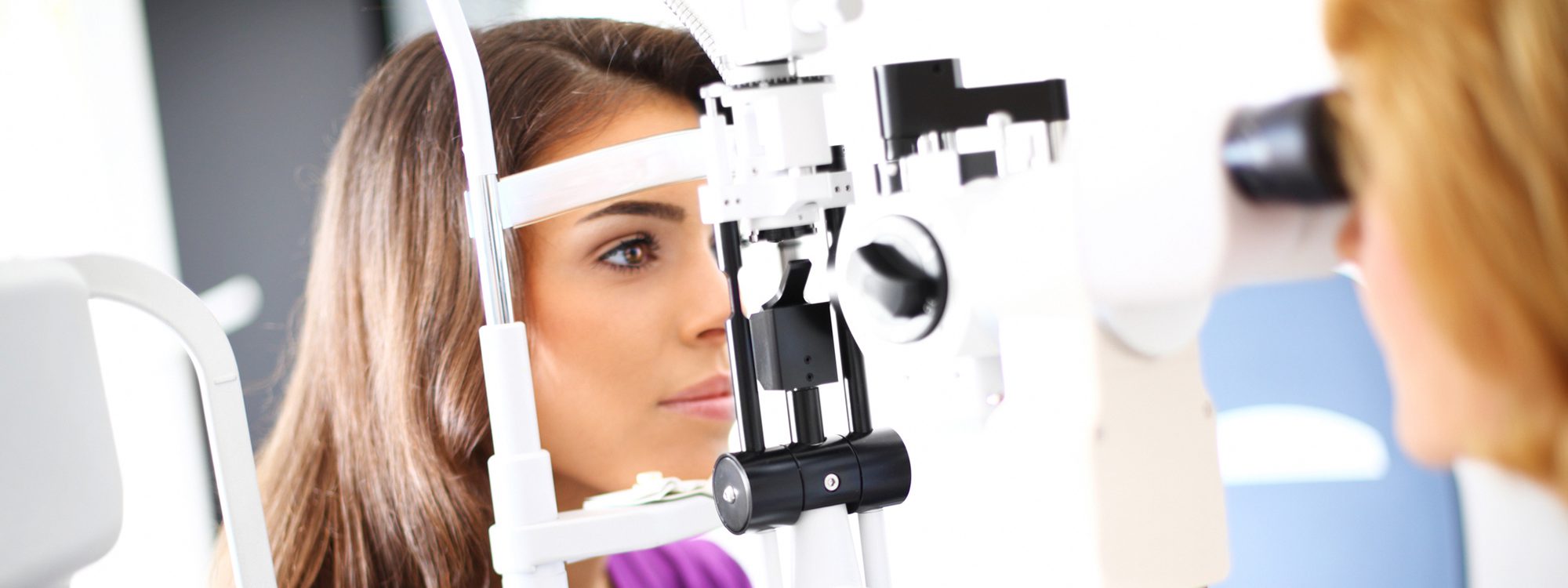
Uncontrolled hypertension is implicated in heart disease, stroke, liver disease and kidney disease; all these are problems of the entire body. What most people don’t think about is that whatever is happening elsewhere in the body is also happening within the eyes. Conversely, changes seen within the eye are also occurring elsewhere. The back of the eye is the only place within the body that blood vessels may be directly viewed without cutting into other tissues, so if a physician sees circulatory problems in the eyes, these problems will also be present in other parts of the body. For this reason, many patients initially learn of their hypertension because their eyecare practitioner examines the back of the eye through dilated pupils.
Circulation to the retina comes from the choroid, a deeply vascular tissue located between the retina and the sclera (the white of the eye) and from the central retinal arteries. All oxygen and nutrients needed by the retina must come from one of these sources.
Causes of Hypertension
There are several causes and conditions that can lead to hypertension, which include
- Kidney disease
- Obesity
- Stress
- Poor diet
- High cholesterol
- Sedentary lifestyle
- Heredity
- Smoking
- Aging
- Alcohol abuse
Retinal Changes
The retina is the light-sensitive layer of tissue that lines the inside of the eye and is made up of nerve fibers that react to light; its main purpose is to send visual impulses through the optic nerve to the brain. Damage to the retina and to the optic nerve from hypertension is caused by interference with the flow of oxygen and necessary nutrients. The amount of damage is largely dependent on the level of high blood pressure and how long it has been present without treatment.
Increased pressure within the arteries causes their walls to thicken, and make them appear as
if they were made of silver or copper wire, because the thickened walls reflect more light back to the examiner. The vessels may also have focal closures, leading to the formation of what are called cotton-wool spots, which appear as small tufts of fluffy cotton, and small superficial hemorrhages. Over time, the arteries may develop tortuosity, or increased curving and bending. As the small arteries continue to harden, they may constrict the veins at locations where the arteries cross over them.
In more severe cases of hypertensive retinopathy, hard exudates form in the retinal tissue;
these can form a ring-shaped reflection around the macula, referred to as a macular star.
Small areas of retinal layer separation can lead to fluid collection and detachment. Dot-and-
blot hemorrhages and flame-shaped hemorrhages can also occur, which are named for their
appearance. The optic nerve head can become swollen, a condition known as papilledema.
Blood pressure levels high enough to cause papilledema and macular star are described as
malignant hypertension.
In more severe cases of hypertensive retinopathy, hard exudates form in the retinal tissue; these can form a ring-shaped reflection around the macula, referred to as a macular star. Small areas of retinal layer separation can lead to fluid collection and detachment. Dot-and-blot hemorrhages and flame-shaped hemorrhages can also occur, which are named for their appearance. The optic nerve head can become swollen, a condition known as papilledema. Blood pressure levels high enough to cause papilledema and macular star are described as malignant hypertension.
Most of these retinal changes, except the most extreme, resolve themselves when the elevated blood pressure is treated and controlled. It is important to note, however, that most of them, like hypertension itself, have no symptoms.
Treatment
Treatment of hypertensive retinopathy involves treating the systemic hypertension causing it. Changes in diet, activity level, smoking cessation and adding medication may be used. A strategy combining two or more of these may be needed to reach a goal of blood pressure under 140/90.
When papilledema and a macular star are present, with extremely elevated blood pressure (for example, 250/150) the patient should be considered to be in a medical crisis and needs immediate transport to a hospital emergency department, preferably by ambulance.
Without symptoms, hypertension can often cause enormous damage before it is diagnosed and treated. One of the best ways to assure that this does not happen is to have regular physical and vision examinations. Ocular effects of hypertension are largely reversible once the hypertension is controlled; however, once damage to the optic nerve and macula occur, there may be a long term reduction in vision.
Regular monitoring of blood pressure is important, and some patients may want to consider getting their own apparatus for doing so. It is often helpful for the physician to have a record of readings done at various times of day, or over a length of time.