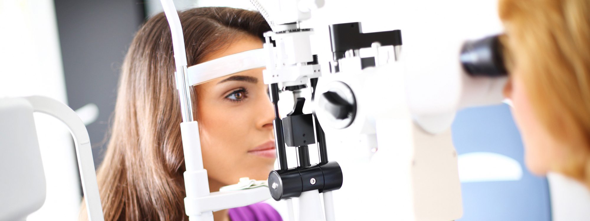
Heidelberg Retina Tomograph (HRT)
Most people equate the eye disease called glaucoma with high eye pressure. It is important to understand that glaucoma is not simply a disease of high intra-ocular pressure, but rather, a more complex disease of the optic nerve. In the early stages of glaucoma there are no symptoms and understandably any condition that lacks symptoms has great potential risk. In glaucoma the vision conductive rim of the optic nerve, called the nerve-fiber layer, is gradually damaged resulting in a lessening of visual information leaving the eye to the brain. We now understand that measurement of eye pressure is only one small part of the data collected for doctors to evaluate the total picture in regard to glaucoma. Up until now the tests utilized to diagnose glaucoma and establish the need for treatment was based on eye pressure and documented vision loss and a diagnosis was only possible after vision loss had already taken place.
Recent developments in computer imaging technology now allow us to image the sensitive nerve-fiber layer of the optic nerve utilizing a three-dimensional cross-section view. The Heidelberg Retinal Tomograph (HRT) is a system that combines a laser-scanning camera and specialized software that evaluates the optic nerve. For the first time this revolutionary technology allows us to understand the progression of optic nerve involvement in regard to glaucoma and other eye conditions long before irreversible vision loss takes place. Tomography utilizes real-time information of the living eye for immediate study and analysis. In addition, each captured image is compared utilizing a normal outcomes database of a patient’s age. It has been shown that 3-D measurements of the optic nerve head are far superior to conventional examination methods.
The HRT works something like an ultrasound, but rather than sound, the process utilizes reflected light and converts the layered image into an enhanced color image. The HRT exam takes just a few minutes and it is a painless non-invasive test. Usually dilation of the eye is not necessary. This technology allows us to more accurately follow disease progression and treatment options and has now become the standard of care for patients with documented cautions, and/or, a strong family history of glaucoma. The key to controlling glaucoma is catching it early. The best way to prevent vision loss from glaucoma is to know your risk factors and to have an eye examination at appropriate intervals.


