Open Angle Glaucoma
Contents |
Open-Angle Glaucoma
Glaucoma is caused by a number of different eye diseases that in most cases produce increased pressure within the eye. This elevated pressure is caused by a backup of fluid in the eye. Over time
it causes damage to the optic nerve and can result in blindness . Open-angle glaucoma the most common form of glaucoma affects over 3 million North Americans – half of whom don t know they have it. Early detection diagnosis and treatment can help prevent serious vision loss and blindness.
Think of your eye as a sink in which the faucet is always running and the drain is always open. The aqueous humor is constantly circulating through the anterior chamber. It is produced by a tiny gland called the ciliary body that is situated behind the iris. It flows between the iris and the lens
and after nourishing the cornea and lens flows out through a very tiny spongy tissue called the trabecular meshwork. The trabecular meshwork is situated in the angle where the iris and cornea meet. When the drain becomes clogged aqueous cannot leave the eye as quickly as it is produced
causing the fluid to back up. But because the eye is a closed compartment the sink doesn t overflow and instead the backed up fluid causes increased pressure to build up within the eye. This is known as open-angle glaucoma.
To understand how this increased pressure affects the eye think of the eye as a balloon. When too much air is blown into the balloon the pressure builds causing it to pop. But the eye is too strong to pop. Instead it gives at the weakest point which is the site in the sclera at which the optic nerve leaves the eye.
The optic nerve is the part of the eye that carries visual information to the brain. It is made up of over one million nerve cells. When the pressure in the eye builds the nerve cells become compressed causing them to become damaged and to eventually die. The death of these cells results in permanent visual loss.
There are usually no symptoms associated with the early stages of open-angle glaucoma. The pressure in the eye slowly rises and vision stays normal and no pain is present. As glaucoma remains untreated however peripheral vision is affected. Unfortunately by the time vision is impaired the damage is irreversible.
Who is at Risk?
Early detection and treatment of glaucoma are the only ways to prevent vision impairment and blindness. There are a few conditions related to this disease which tend to put some people at greater risk:
People over the age of 45
People who have a family history of glaucoma
Glaucoma appears to run in families. The tendency for developing glaucoma may be inherited. However
just because someone in your family has glaucoma does not mean that you will necessary develop the disease.
People with abnormally high intraocular pressure (IOP). The aqueous humor provides the necessary pressure to help maintain the shape of the eye. This pressure is called the intaocular pressure (IOP). Abnormally high IOP may increase the risk of glaucoma.
People of African descent.
People who have:
- Diabetes
- High Myopia (nearsightedness)
- Regular long-term steroid/cortisone use
- A previous eye injury
Diagnosing Glaucoma
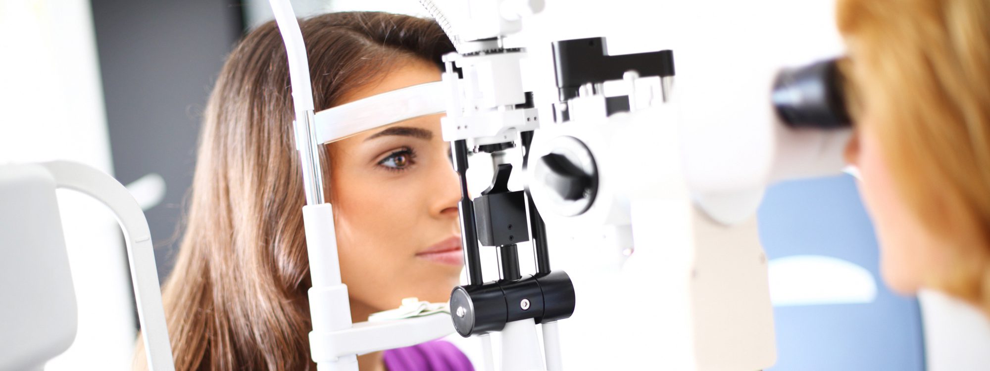
A variety of diagnostic tools aid in testing for glaucoma
The Tonometer — The tonometer measures the pressure in the eye. In applanation tonometry anesthetic drops are used to numb the eye and a small pressure-sensitive device is applied to the eye to measure the intraocular pressure. In air tonometry a puff of air is sent onto the cornea to take the measurement. Since this instrument does not come in direct contact with your eye no anesthetic eye drops are required.
Visual Field Test — The visual field test is an extremely important part of the examination for glaucoma. Glaucoma causes loss of side vision long before central vision becomes damaged and the only way to test side vision is with the visual field test. The visual field is an important measure of the extent of damage to the optic nerve from increased IOP. In glaucoma once a sufficient number of nerve cells are destroyed blind spots begin to form in the peripheral field of vision.
Ophthalmoscopy — Using an instrument called an ophthalmoscope the optic nerve at the back of the eye can be observed. The optic nerve is best observed when drops are put into the eyes to enlarge (dilate) the pupils. Left untreated elevated pressure in the eye can damage the optic nerve.
Treatment of Glaucoma
Although glaucoma can t be cured there are effective treatments to control it. Glaucoma can be treated with prescription eyedrops laser surgery eye operations or a combination of methods. The whole prupose of treatment is to prevent further loss of vision. This is imperitave as loss of vision due to glaucoma is irreversible. Keeping the intraocular pressure under control is the key to preventing loss of vision from glaucoma.

